
Introduction
This case study presents the clinical details and imaging observations of a 49-year-old male patient who complained of left side abdominal pain accompanied by fever. Through a comprehensive radiological examination, including CT scans, we aim to delineate the underlying pathology, which includes a left vesico-ureteric junction calculus leading to mild hydroureteronephrosis, acute pyelo-ureteritis, and associated perinephric findings.
Clinical Details
- Patient: 49-year-old male
- Complaint: Left side abdominal pain with fever
Observations
Liver
- Condition: Normal in size and uniform density.
- Details: No focal lesions or abnormal enhancements observed.
- Biliary Radicles: Normal.
- Portal and Hepatic Veins: Normal.
Common Bile Duct (CBD)
- Condition: Not dilated.
Gallbladder (GB)
- Findings: No radio-opaque calculi.
Pancreas
- Condition: Normal in size and density.
- Details: No calcifications, masses, or peripancreatic fluid collections.
- Pancreatic Duct: Not dilated.
Spleen
- Condition: Normal in size and density.
Right Kidney
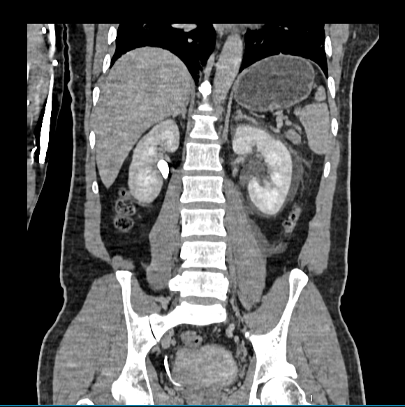
- Size: Measures 10 x 4.9 cm.
- Pelvicalyceal System: Not dilated.
- Enhancement: Homogeneous enhancement during the nephrogenic phase.
- Ureter: Not dilated; no calculus seen.
- Vesico-Ureteric Junction: Appears normal.
Left Kidney
- Size: Measures 11 x 4.7 cm.
- Pelvicalyceal System: Not dilated.
- Enhancement: Homogeneous enhancement during the nephrogenic phase.
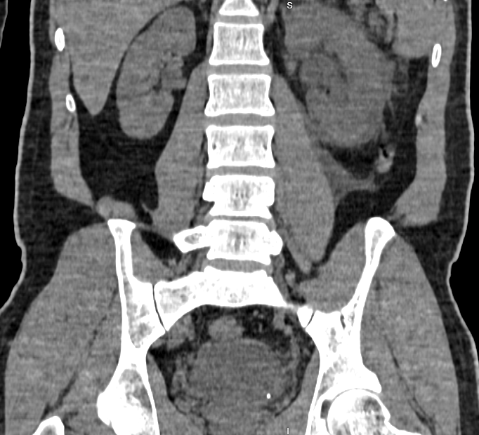
- Calculus: Small calculus of size approximately 3.2 x 3 mm at the left vesico-ureteric junction, causing mild hydroureteronephrosis.
- Urothelial Thickening: Hyperenhancement involving renal pelvis and ureter, suggestive of acute pyelo-ureteritis.
- Perinephric Findings: Fat stranding, Gerota’s fascia thickening, and mild perinephric fluid collection.
- Ureter: Left ureter is not opacified on the delayed scan.
Other Findings
- Retroperitoneum: No mass or lymphadenopathy observed.
- Peritoneal Cavity: No free fluid seen.
- Bowel: No obvious wall thickening or dilatation; no evidence of acute appendicitis.
- Bladder: Normal, with no evidence of diverticulum or calculus.
- Prostate: Normal.
- Dorsolumbar Spine: Visualized spine shows degenerative changes.
- Lung Fields: Visualized lung fields show dependent opacities in both lower lobes.
- Abdominal Wall: Appears normal.
Impression
- Left Vesico-Ureteric Junction Calculus: Causing mild hydroureteronephrosis.
- Left Perinephric Fat Stranding: Gerota’s fascia thickening and mild perinephric fluid collection.
- Left Acute Pyelo-Ureteritis