Patient Detail : 57 Yr / Female
History: Complain of pain abdomen, vomiting and loss of appetite since 3 days.
Clinical Observations:
- Liver measures 11.2 cm in craniocaudal span, shows normal attenuation& outline. No focal lesion noted. No IHBRD seen.
- Gall bladder is not visualized. Surgical clips are seen in GB fossa ( cholecystectomy)
- Spleen is normal in size measuring 8.8 cm, shows normal attenuation. No focal lesion noted.
- Pancreas is normal in bulk and attenuation. No focal lesion seen. MPD is not dilated.
- Right kidney measures 8.7 x 3.9 cm, normal in size and show normal outline and nephrographic density. No calculi / hydronephrosis. No perinephric fat stranding / collection noted.
- Right ureter is normal in course and caliber.
- Left kidney measures 9.6 x 4.4 cm , normal in size and show normal outline and nephrographic density. No calculi / hydronephrosis. No perinephric fat stranding / collection noted.
- Left ureter is normal in course and caliber.
- Bilateral adrenal glands are normal in size and attenuation. No mass lesion is seen.
- A large sigmoid colon diverticulum is seen.
- Rest of the bowel loops are normal.
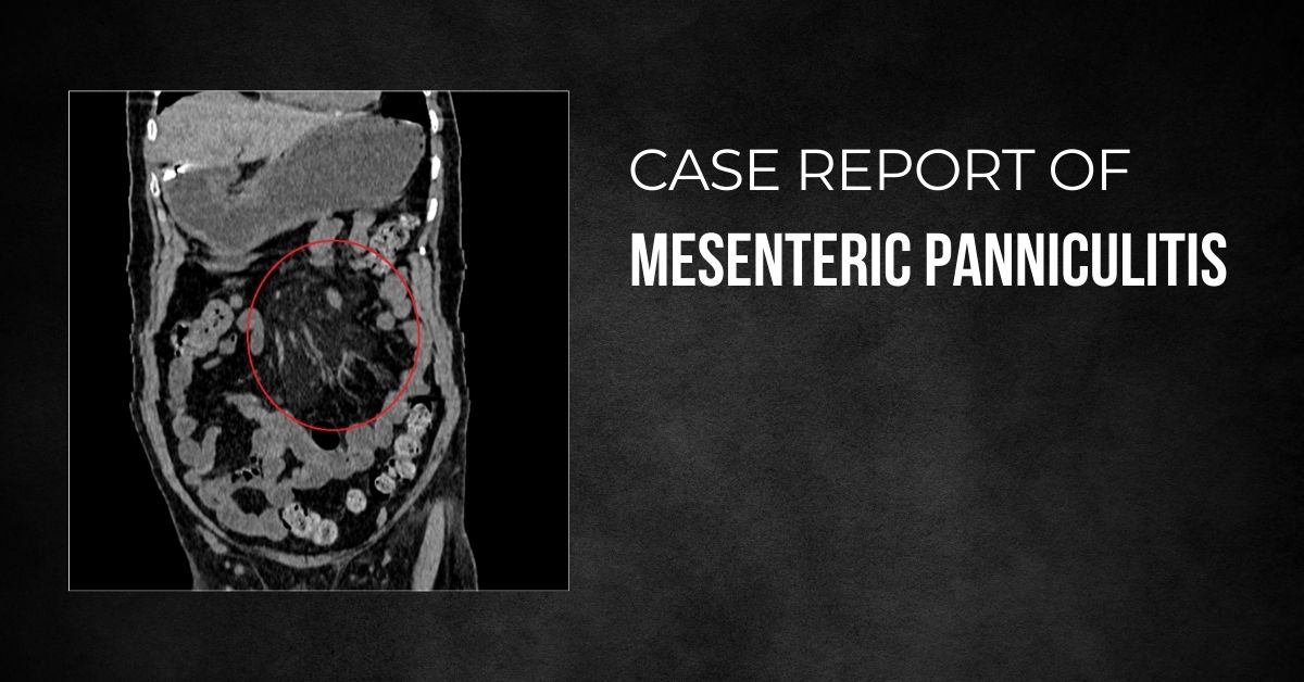
- Inhomogeneouslyhyperdense mesenteric fat lesion with misty mesentery is seen surrounding the superior mesenteric vessels and their branches within the root of the small bowel mesentery. Few small mesenteric lymph nodes are seen. CT features are suggestive mesenteric panniculitis.
- No ascites is seen.
- Urinary bladder is not distended. Foleys catheter seen in situ.
- Uterus and both adnexa are unremarkable.
- Visualised bilateral lungs are normal.
- Visualized spine show degenerative changes.
- T11 vertebral body haemangioma is seen.
- Abdominal aorta and its branches show atherosclerotic changes.
Impression:
NCCT abdomen features are suggestive of –
Mesenteric panniculitis.
Large Sigmoid colon diverticulum.
T11 vertebral body haemangioma.