
Patient details:
55 years /Female Complain of pain abdomen from 15 days
Observations:
- Liver measures 13.1 cm in craniocaudal span and shows normal outline and attenuation. A small calcific foci is seen in segment VI of liver- likely old healed granuloma.
- Gall bladder is distended and is normal. Common bile duct is not dilated. No intrahepatic biliary duct dilatation is seen.
- Spleen measures 8.6 cm in craniocaudal span and shows normal attenuation. No focal lesion seen.
- Pancreas is normal in size and attenuation. No focal lesion seen. Main pancreatic duct is not dilated.
- Bilateral kidneys are normal in size and show normal outline and nephrographic density. Bilateral pelvi-calycealsystem are compact. Bilateral ureters are not dilated.
- Bilateral adrenal glands are normal in size and attenuation. No mass lesion is seen.
- The bowel loops are normal in caliber and mural thickness. No mass lesion seen.
- Aorta and its branches, IVC and its tributaries and spleno-portal axis are normal.
- No significant mesenteric or retroperitoneal lymphadenopathy is seen.
- No ascites is seen.
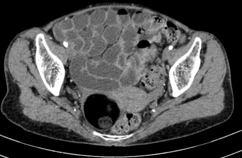
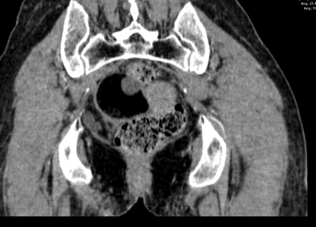
- Urinary bladder is distended and normal.
- Uterus and left ovary is unremarkable.
- There is a well defined right adnexal mass which contains mostly fat with two well-defined non enhancing soft tissue nodule along posterior wall and anteromedial wall. The mass measures approximately 5.1 x 4.4 x4.0 cm.
- No lymphadenopathy is seen.
- CECT features are suggestive of right ovarian dermoid cyst
- Visualized bilateral lungs and bones are unremarkable.
Impression:
CT findings are suggestive of Right ovarian dermoid cyst.
Advice- Histopathological correlation.